

Cross section of human skin under microscope view for education in laboratory.
Коллекция по умолчанию
Коллекция по умолчанию
Создать новую
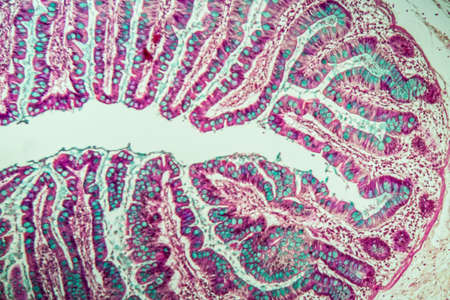
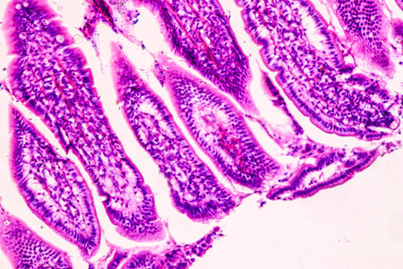
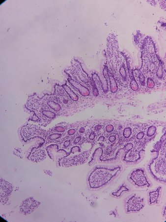
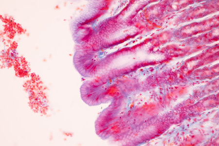
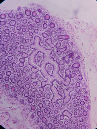
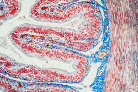
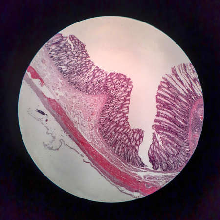
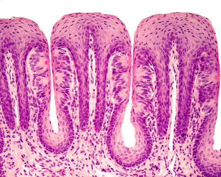
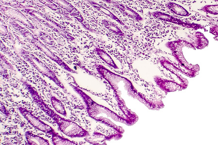
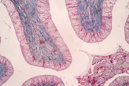
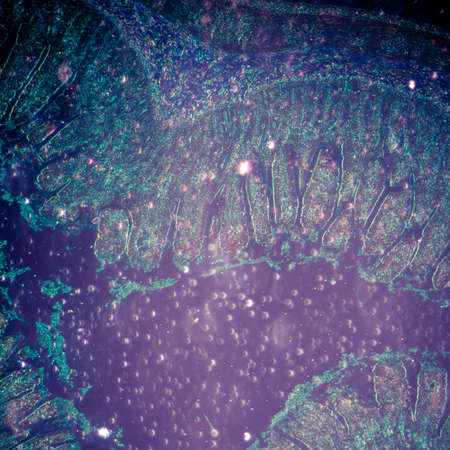
Small intestine with villi under the microscope 100x
Коллекция по умолчанию
Коллекция по умолчанию
Создать новую

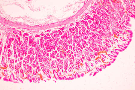
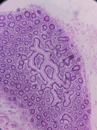
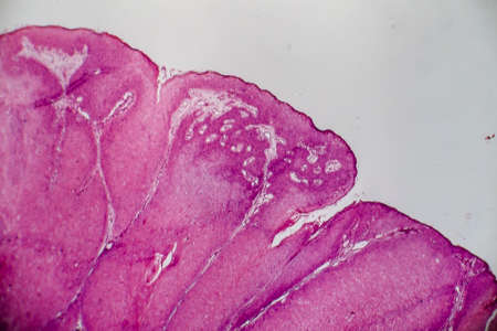
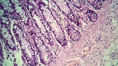
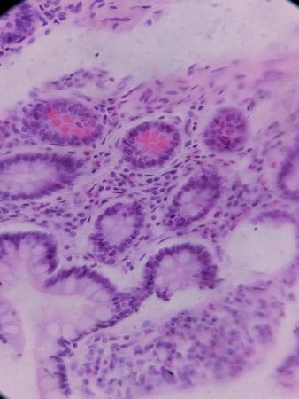
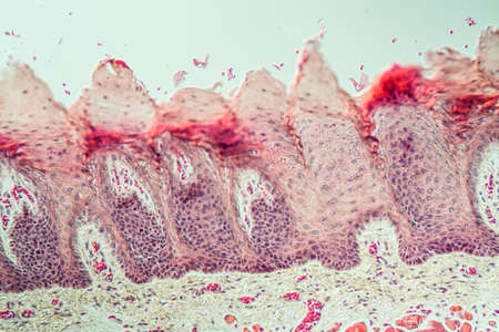
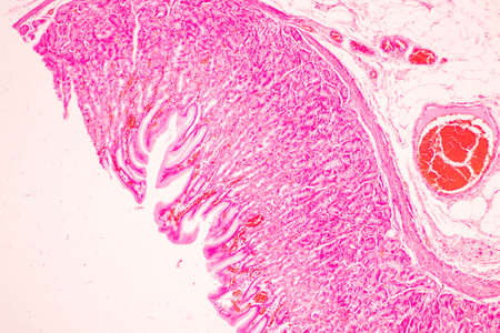
Stomach tissue under the microscope 100x
Коллекция по умолчанию
Коллекция по умолчанию
Создать новую
Condyloma acuminatum, also known as genital warts. Light micrograph, photo under microscope
Коллекция по умолчанию
Коллекция по умолчанию
Создать новую

Ovarian cancer, light micrograph, photo under microscope. Photograph shows a fragment of a cancerous tumor in the female ovary. Selective focus
Коллекция по умолчанию
Коллекция по умолчанию
Создать новую
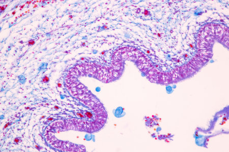
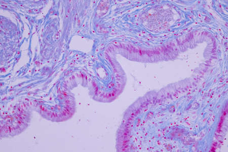
Columnar epithelium of human gall bladder under the microscope in Lab.
Коллекция по умолчанию
Коллекция по умолчанию
Создать новую
Pathology and Histology Tissue of Mammals under microscope.
Коллекция по умолчанию
Коллекция по умолчанию
Создать новую
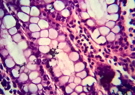
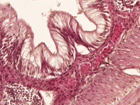
Tissue of Stomach Human under the microscope in Lab.
Коллекция по умолчанию
Коллекция по умолчанию
Создать новую
Education anatomy and Histological sample of Human under the microscope.
Коллекция по умолчанию
Коллекция по умолчанию
Создать новую
Stomach tissue under the microscope 100x
Коллекция по умолчанию
Коллекция по умолчанию
Создать новую
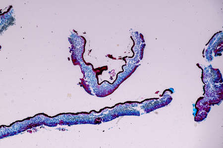
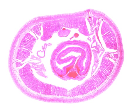
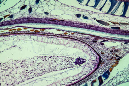
Slow worm histology bowel transverse 100x
Коллекция по умолчанию
Коллекция по умолчанию
Создать новую
Histopathology of cholera under microscope view for education.
Коллекция по умолчанию
Коллекция по умолчанию
Создать новую

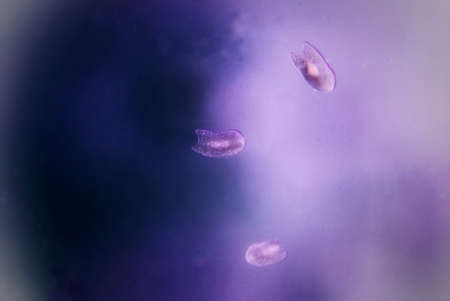
Translucent jellyfish swim elegantly in dark waters, displaying stunning forms and luminescent features, captivating viewers with their serene motion.
Коллекция по умолчанию
Коллекция по умолчанию
Создать новую
Pancreas cancer cell under microscope view for medical education.
Коллекция по умолчанию
Коллекция по умолчанию
Создать новую
Bacteria cells under microscope, 3D illustration.
Коллекция по умолчанию
Коллекция по умолчанию
Создать новую
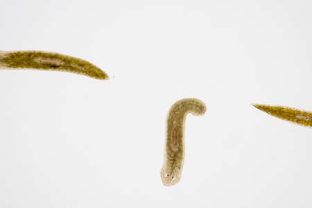
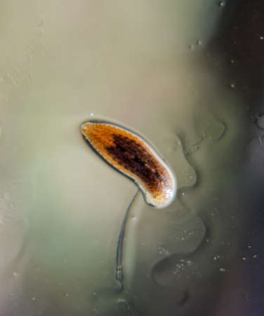
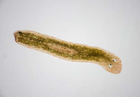
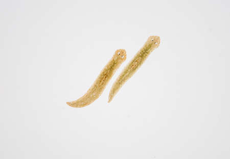
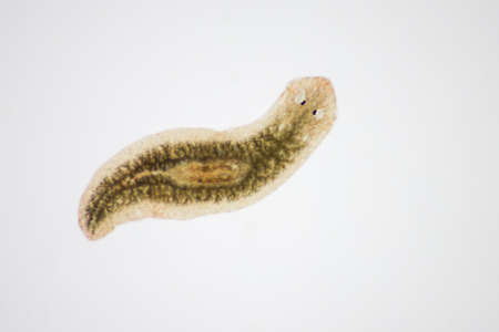
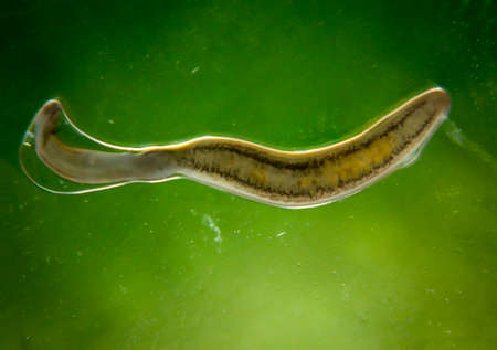
Planarian parasite (flatworm) under microscope view.
Коллекция по умолчанию
Коллекция по умолчанию
Создать новую
Histopathology of human under microscope view for education in laboratory.
Коллекция по умолчанию
Коллекция по умолчанию
Создать новую

Cross section of human body under microscope view for education in laboratory.
Коллекция по умолчанию
Коллекция по умолчанию
Создать новую
Condyloma acuminatum, also known as genital warts. Light micrograph, photo under microscope
Коллекция по умолчанию
Коллекция по умолчанию
Создать новую

Histopathology of human under microscope view for education in laboratory.
Коллекция по умолчанию
Коллекция по умолчанию
Создать новую
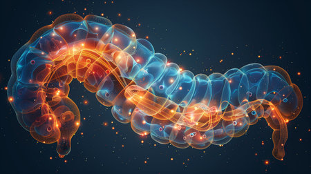
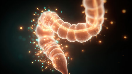
A Digital Rendering of a Human Intestine With Glowing Particles
Коллекция по умолчанию
Коллекция по умолчанию
Создать новую

Histological Uterus human, Uterine tube human, Placenta human and Umbilical cord Human under the microscope for education.
Коллекция по умолчанию
Коллекция по умолчанию
Создать новую
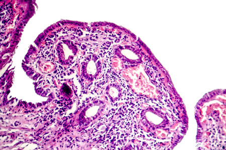
Intestinal polypoid adenoma, light micrograph, photo under microscope
Коллекция по умолчанию
Коллекция по умолчанию
Создать новую

Abstract science background- pyloric division of the stomach of the dog. Cell biology
Коллекция по умолчанию
Коллекция по умолчанию
Создать новую

Endometriosis, a disorder in which cells similar to those in the endometrium grow outside the uterus. Light micrograph, photo under microscope
Коллекция по умолчанию
Коллекция по умолчанию
Создать новую

Colon polyp, one of the largest polyps
Коллекция по умолчанию
Коллекция по умолчанию
Создать новую
Bacillary dysentery, light micrograph, photo under microscope showing presence of bacteria and accumulation of inflammatory cells in intestinal epithelium
Коллекция по умолчанию
Коллекция по умолчанию
Создать новую
Discover a captivating 3D visualization of mitochondria, showcasing vibrant colors and a glowing effect. Perfect for scientific, educational, and artistic purposes.
Коллекция по умолчанию
Коллекция по умолчанию
Создать новую
Extreme Close up of microscopic kidney Bowman's Capsule and Glomerulus
Коллекция по умолчанию
Коллекция по умолчанию
Создать новую

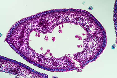
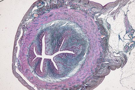
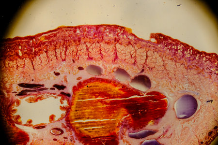
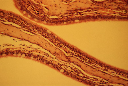
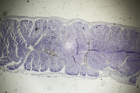
Earthworm histology cross section 10th segment 100x
Коллекция по умолчанию
Коллекция по умолчанию
Создать новую

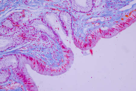
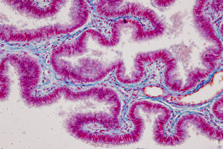
Columnar epithelium of human gall bladder under the microscope in Lab.
Коллекция по умолчанию
Коллекция по умолчанию
Создать новую
Leech on the glass. Bloodsucking animal. subclass of ringworms from the belt-type class. Hirudotherapy.
Коллекция по умолчанию
Коллекция по умолчанию
Создать новую

Columnar epithelium of human gall bladder under the microscope in Lab.
Коллекция по умолчанию
Коллекция по умолчанию
Создать новую
Microscopic view of tissue with purple staining, showing cellular structures and patterns, possibly from a biological or medical sample
Коллекция по умолчанию
Коллекция по умолчанию
Создать новую
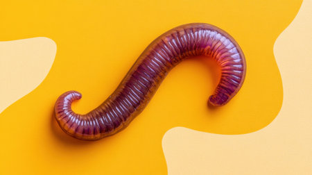
A striking image of a colorful purple worm set against a vibrant yellow background. Ideal for creative projects, educational materials, or graphic design.
Коллекция по умолчанию
Коллекция по умолчанию
Создать новую
Histopathology of human liver under microscope view for medical education.
Коллекция по умолчанию
Коллекция по умолчанию
Создать новую
Watercolor abstract. Red paint texture of watercolor pattern or splash ink stain for design isolated on water color background
Коллекция по умолчанию
Коллекция по умолчанию
Создать новую

A deep stunning-sea siphonophore drifting gracefully in the ocean depths
Коллекция по умолчанию
Коллекция по умолчанию
Создать новую
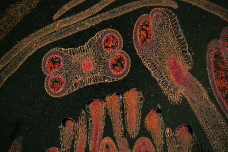
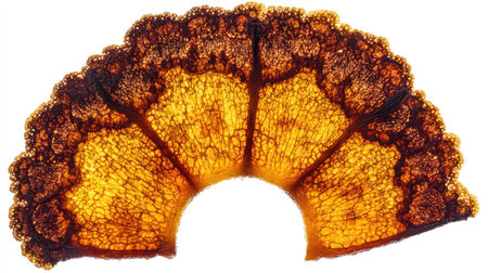
Strawberry flower bud in cross section 100x
Коллекция по умолчанию
Коллекция по умолчанию
Создать новую
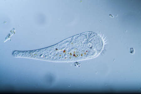
Trumpet animal as a microscopic plankton animal in drops of water
Коллекция по умолчанию
Коллекция по умолчанию
Создать новую
Columnar epithelium of human gall bladder under the microscope in Lab.
Коллекция по умолчанию
Коллекция по умолчанию
Создать новую
Abstract background of acrylic paint in pink and blue tones
Коллекция по умолчанию
Коллекция по умолчанию
Создать новую
Characteristics of Lichen, hyphae and Symbiotic algae under the microscope for education.
Коллекция по умолчанию
Коллекция по умолчанию
Создать новую
Heather leaf cross section under the microscope, 200x
Коллекция по умолчанию
Коллекция по умолчанию
Создать новую
Planarian parasite (flatworm) under microscope view.
Коллекция по умолчанию
Коллекция по умолчанию
Создать новую
black white abstract acrylic painting color texture on white paper background by using rorschach inkblot method
Коллекция по умолчанию
Коллекция по умолчанию
Создать новую
Small intestine tissue under the microscope 200x
Коллекция по умолчанию
Коллекция по умолчанию
Создать новую
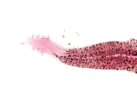
Earthworm under the microscope, background Lumbricidae
Коллекция по умолчанию
Коллекция по умолчанию
Создать новую
Planarian parasite (flatworm) under microscope view.
Коллекция по умолчанию
Коллекция по умолчанию
Создать новую
Pathology and Histology Tissue of Mammals under microscope.
Коллекция по умолчанию
Коллекция по умолчанию
Создать новую
Rare image of Ghost flatworm - Maricola (Planarian) triclad flatworms in reef aquarium glass
Коллекция по умолчанию
Коллекция по умолчанию
Создать новую
Bacillary dysentery, light micrograph, photo under microscope showing presence of bacteria and accumulation of inflammatory cells in intestinal epithelium
Коллекция по умолчанию
Коллекция по умолчанию
Создать новую
Chronic cholecystitis, light micrograph, photo under microscope showing fibrosis and muscular hypertrophy of gallbladder wall, entrapped epithelial crypts, foamy macrophages
Коллекция по умолчанию
Коллекция по умолчанию
Создать новую
Planarian parasite (flatworm) under microscope view.
Коллекция по умолчанию
Коллекция по умолчанию
Создать новую
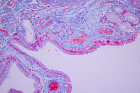
Showing Light micrograph of the Trachea, Thymus, Parathyroid gland and Tonsil human under the microscope for education in the laboratory.
Коллекция по умолчанию
Коллекция по умолчанию
Создать новую
Bacillary dysentery, light micrograph, photo under microscope showing presence of bacteria and accumulation of inflammatory cells in intestinal epithelium
Коллекция по умолчанию
Коллекция по умолчанию
Создать новую

Endometriosis, a disorder in which cells similar to those in the endometrium grow outside the uterus. Light micrograph, photo under microscope
Коллекция по умолчанию
Коллекция по умолчанию
Создать новую
Microscopic view of tissue with purple staining, showcasing cellular structures and patterns
Коллекция по умолчанию
Коллекция по умолчанию
Создать новую
science aquaculture fish parasite hook clip worm micrograph
Коллекция по умолчанию
Коллекция по умолчанию
Создать новую
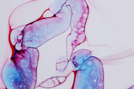
Leech cross section showing internal anatomical structures stained
Коллекция по умолчанию
Коллекция по умолчанию
Создать новую
Taste buds in foliate tongue papillae. Many of them show the taste or gustatory pore. Hematoxylin & eosin stain.
Коллекция по умолчанию
Коллекция по умолчанию
Создать новую

Education anatomy and Histological sample Spinal cord Tissue under the microscope.
Коллекция по умолчанию
Коллекция по умолчанию
Создать новую

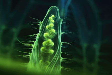
Proglottid (body unit) of tapeworm Taenia saginata, 3D illustration. A flatworm parasitizing animal and human intestine. Proglottid contains uterus with 12-30 primary lateral branches filled with eggs
Коллекция по умолчанию
Коллекция по умолчанию
Создать новую
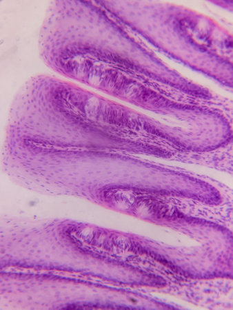
Tongue Tissue with taste buds across 200x
Коллекция по умолчанию
Коллекция по умолчанию
Создать новую
A glowing, glowing, glowing, glowing, glowing, glowing, glowing, glowing, glowing, glowing, glowing, glowing, glowing, glowing, glowing, glowing, glowing, glowing, glowing, glowing, glowing, glowing,
Коллекция по умолчанию
Коллекция по умолчанию
Создать новую
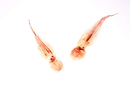
Dried octopus isolated on a white backgrouund.
Коллекция по умолчанию
Коллекция по умолчанию
Создать новую
Nasal cavity
Коллекция по умолчанию
Коллекция по умолчанию
Создать новую
Large intestine with diverticular tissue under the microscope 100x
Коллекция по умолчанию
Коллекция по умолчанию
Создать новую

Planarian parasite (flatworm) under microscope view.
Коллекция по умолчанию
Коллекция по умолчанию
Создать новую
Intestine animal tissue under microscope view. histology of intestine.
Коллекция по умолчанию
Коллекция по умолчанию
Создать новую

Paper cutout of small intestine on light green background, top view
Коллекция по умолчанию
Коллекция по умолчанию
Создать новую

Paramecium caudatum is a genus of unicellular ciliated protozoan and Bacterium under the microscope.
Коллекция по умолчанию
Коллекция по умолчанию
Создать новую
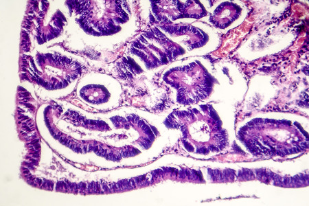
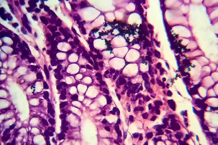
Intestinal metaplasia of stomach, light micrograph, photo under microscope
Коллекция по умолчанию
Коллекция по умолчанию
Создать новую
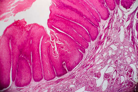
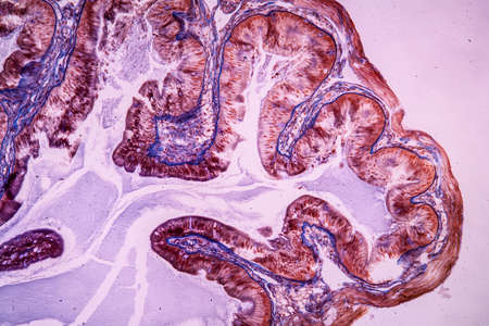
Tissue of Stomach Human under the microscope in Lab.
Коллекция по умолчанию
Коллекция по умолчанию
Создать новую
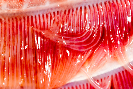
pike fish gills. super macro
Коллекция по умолчанию
Коллекция по умолчанию
Создать новую
Concept of health care, stomach and intestinal problems
Коллекция по умолчанию
Коллекция по умолчанию
Создать новую

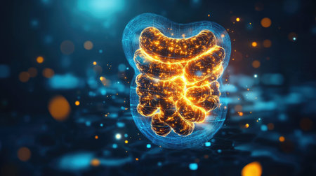
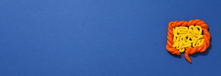
Three-dimensional wishing model of human intestines on a blue background, 3D rendering
Коллекция по умолчанию
Коллекция по умолчанию
Создать новую
Histological Uterus human, Uterine tube human, Placenta human and Umbilical cord Human under the microscope for education.
Коллекция по умолчанию
Коллекция по умолчанию
Создать новую
Proglottid (body unit) of tapeworm Taenia saginata, 3D illustration. A flatworm parasitizing animal and human intestine. Proglottid contains uterus with 12-30 primary lateral branches filled with eggs
Коллекция по умолчанию
Коллекция по умолчанию
Создать новую

Backgrounds of Characteristics Tissue of Stomach Human, Small intestine Human, Pancreas Human and Large intestine Human under the microscope in Lab.
Коллекция по умолчанию
Коллекция по умолчанию
Создать новую

3D illustration of abstract fractal, digital artwork for creative graphic design
Коллекция по умолчанию
Коллекция по умолчанию
Создать новую
Columnar epithelium of human gall bladder under the microscope in Lab.
Коллекция по умолчанию
Коллекция по умолчанию
Создать новую
Johannes berry fruit cross 100x
Коллекция по умолчанию
Коллекция по умолчанию
Создать новую
An extreme macro of an Asian leech
Коллекция по умолчанию
Коллекция по умолчанию
Создать новую
Pathology and Histology Tissue of Mammals under microscope.
Коллекция по умолчанию
Коллекция по умолчанию
Создать новую
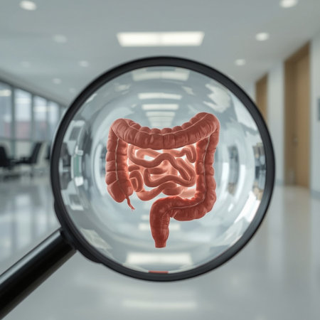
Magnifying glass enlarging large intestine inside hospital building. 3D rendering
Коллекция по умолчанию
Коллекция по умолчанию
Создать новую
Bacillary dysentery, light micrograph, photo under microscope showing presence of bacteria and accumulation of inflammatory cells in intestinal epithelium
Коллекция по умолчанию
Коллекция по умолчанию
Создать новую
Skeletal muscle section under the microscope
Коллекция по умолчанию
Коллекция по умолчанию
Создать новую
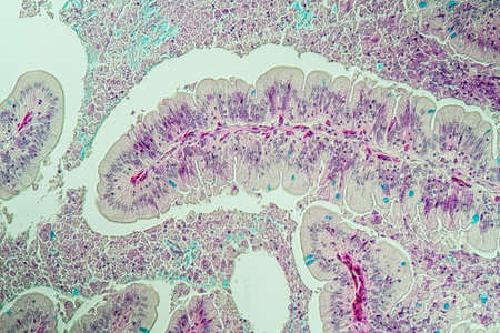
science medical anthropotomy physiology micrograph of small intestinum tenue tissue
Коллекция по умолчанию
Коллекция по умолчанию
Создать новую
euglena magnified against a bright backdrop, created with generative ai
Коллекция по умолчанию
Коллекция по умолчанию
Создать новую

Small and large Intestine 3D Illustration Human Digestive System Anatomy For Medical Concept
Коллекция по умолчанию
Коллекция по умолчанию
Создать новую
Cerebellum and Nerve human under the microscope for education in Lab.
Коллекция по умолчанию
Коллекция по умолчанию
Создать новую
This stunning orange leaf fragment highlights intricate textures and patterns, making it an ideal choice for nature-themed designs and artistic projects.
Коллекция по умолчанию
Коллекция по умолчанию
Создать новую

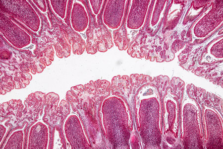
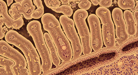
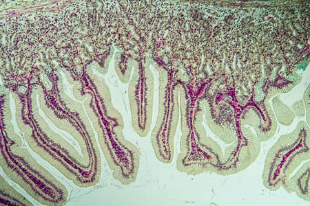
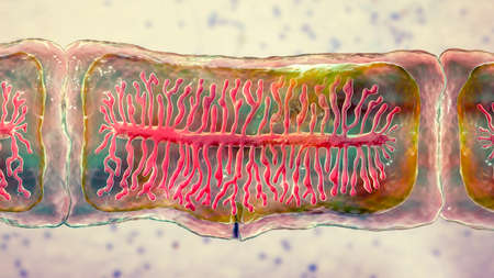
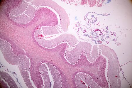
Villi of small intestine, light micrograph, magnification 100x
Коллекция по умолчанию
Коллекция по умолчанию
Создать новую
Microscopic view of tissue showing purple-stained cells and structures, highlighting cellular organization and morphology
Коллекция по умолчанию
Коллекция по умолчанию
Создать новую
Histopathology of intestinal adenoma, light micrograph, photo under microscope
Коллекция по умолчанию
Коллекция по умолчанию
Создать новую
Small sandeel fishes in sea water.
Коллекция по умолчанию
Коллекция по умолчанию
Создать новую

science medical anthropotomy physiology micrograph of small intestinum tenue tissue
Коллекция по умолчанию
Коллекция по умолчанию
Создать новую
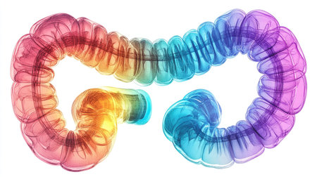
This image features a vibrant and colorful artistic representation of the human intestine, showcasing gradient effects and abstract design elements ideal for educational purposes.
Коллекция по умолчанию
Коллекция по умолчанию
Создать новую
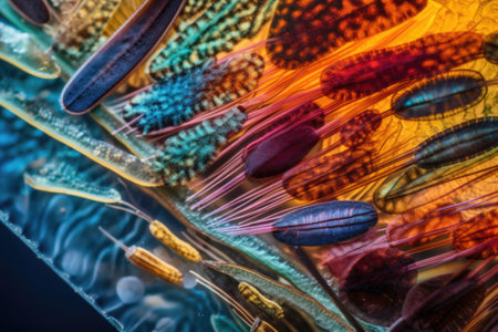
close-up of colorful marine organism on microscope slide, created with generative ai
Коллекция по умолчанию
Коллекция по умолчанию
Создать новую
Legion-Media
Создайте свои проекты на основе качественных стоковых фотографий и видео.


Copyright © Legion-Media.